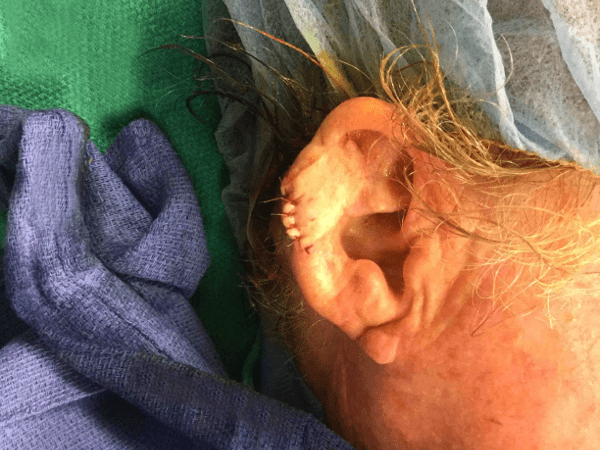
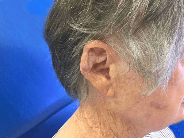
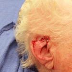
Mohs Ear Patient 09
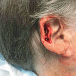
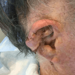
Patient with a history of basal cell carcinoma to the right ear superior helical sulcus. Patient underwent Mohs surgery in two stages resulting in a 3.0 cm x 1.5 cm defect. Patient underwent reconstruction with a pedicle flap.
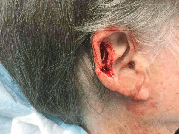
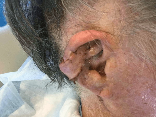
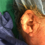
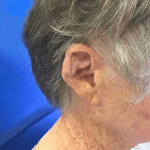
Mohs Ear Patient 04

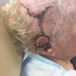
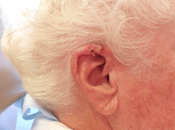
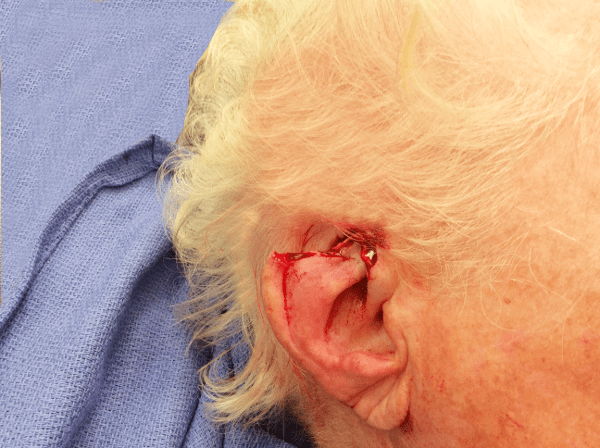
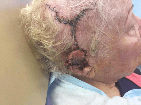
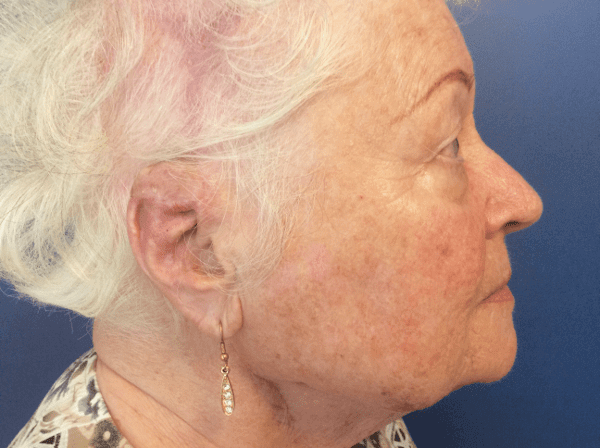
Patient with history of basal cell carcinoma right ear superior helix. Patient underwent Mohs surgery in five stages and reconstruction with pedicled temporal parietal fascia flap, ipsilateral ear cartilage graft and split-thickness skin graft.
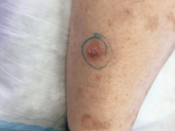
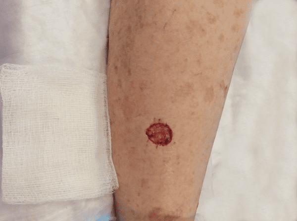
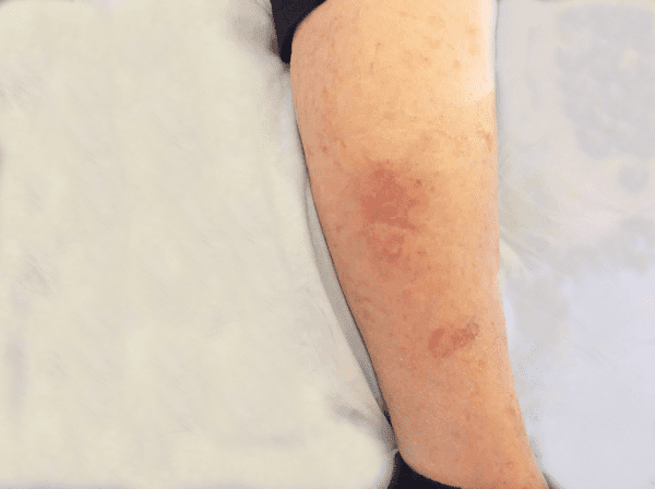
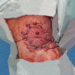
Mohs Leg Patient 01
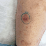
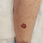
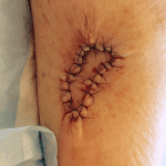
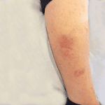
Patient with a history of squamous cell carcinoma to the left lateral mid tibia. Patient underwent Mohs surgery and the 19mm x 17mm defect was reconstructed with a Keystone flap repair. Final picture 5 weeks post operative.
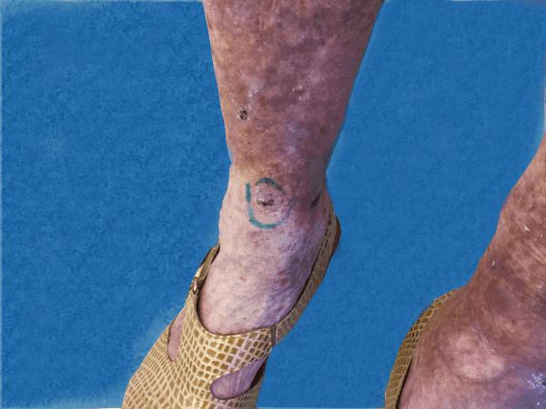
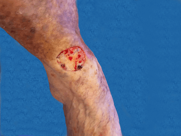
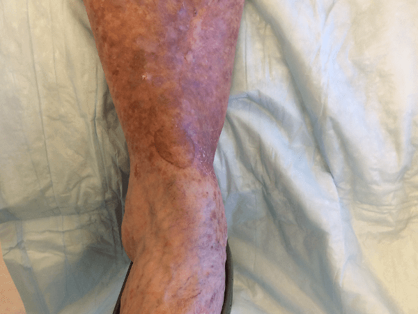
Mohs Leg Patient 02

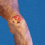
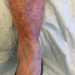
Patient with a history of squamous cell carcinoma above the right foot. Patient underwent Mohs surgery and the 3.0cm x 3.5cm defect was reconstructed with a split thickness skin graft.
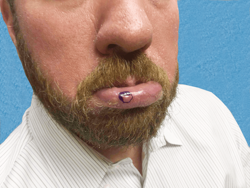
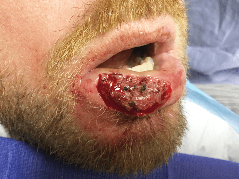
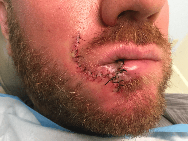
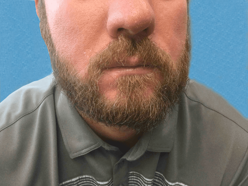
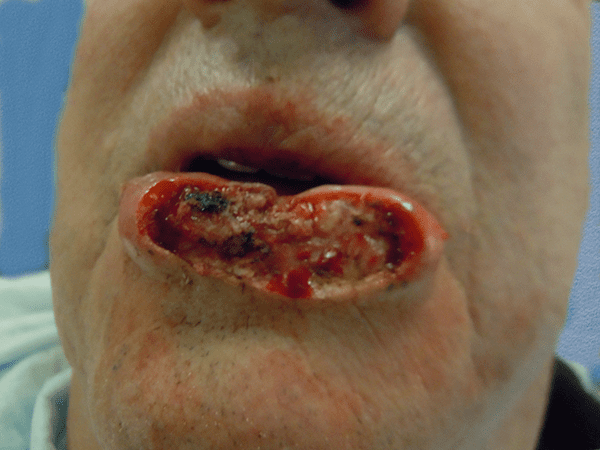
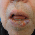
Mohs Lips Patient 04
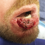
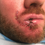
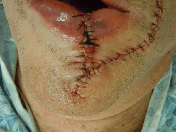
Patient with a history of squamous cell carcinoma to the right medial lower lip. Patient underwent Mohs surgery in seven stages resulting in a 3.0cm x 1.9cm defect. Patient underwent reconstruction with a karapandzic flap closure.
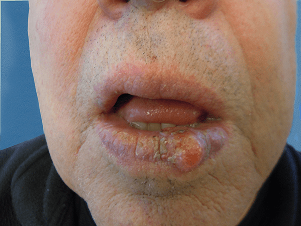
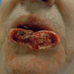
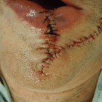
Mohs Lips Patient 01
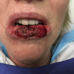

Patient with history of chronic chapping of the lower lip. Diagnosis was squamous cell carcinoma. Patient underwent Mohs surgery with 80% extensive defect of lower lip. Patient underwent bilateral Karapandzic flap reconstruction.
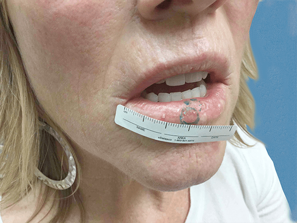
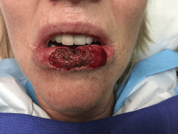
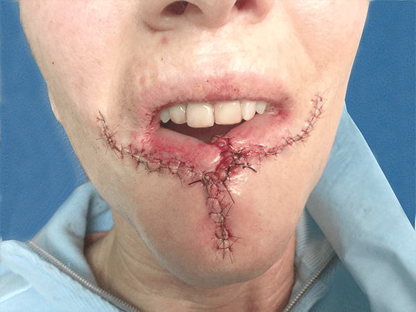
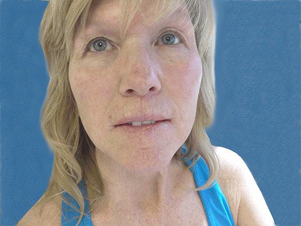
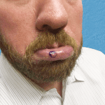
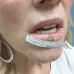
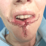
Mohs Lips Patient 02
This is a patient with squamous cell carcinoma of lower lip. Patient underwent Mohs surgery with extensive defect involved 70% of the lip. Patient underwent Karapandzic flap reconstruction.
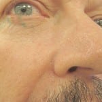
Mohs Nose Patient 21
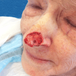

Patient with a history of squamous cell carcinoma to the left lateral nasal tip. Patient underwent Mohs surgery in three stages resulting in a 2.6 cm x 2.0 cm defect. Patient underwent reconstruction with a pedicled nasolabial flap and subsequent division and inset of flap.
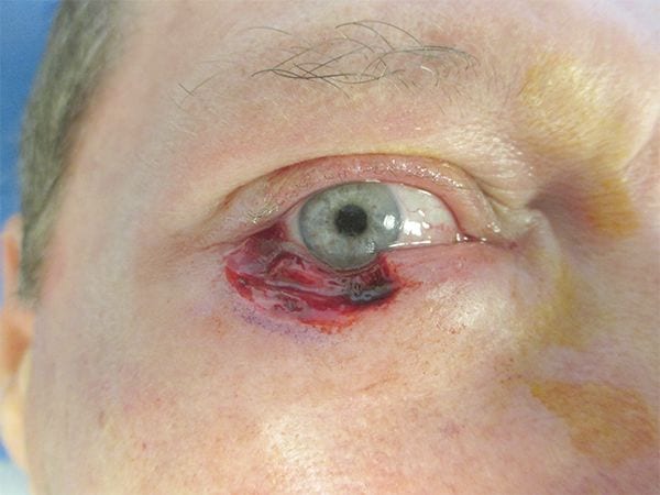
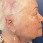
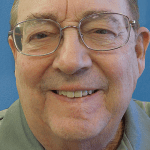
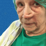
Mohs Eyes Patient 01
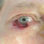


Patient with history of gradually increasing pigmentation on right lateral lower eyelid margin. Patient diagnosed with a melanoma in-situ, underwent resection with clear margins obtained. Patient then underwent right lateral eyelid reconstruction with Hughes tarsoconjunctival flap with full thickness skin graft, and subsequent division.
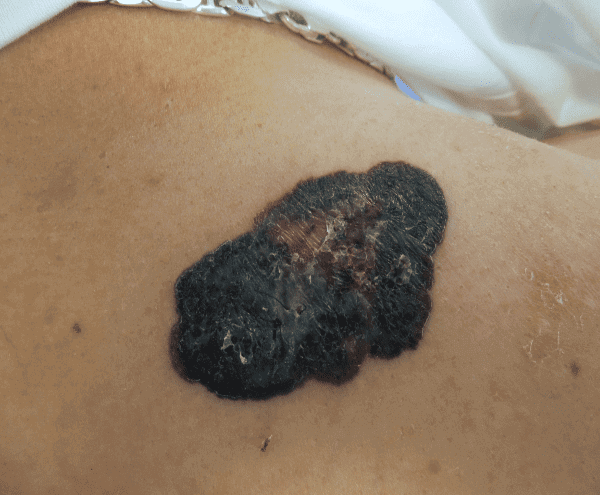
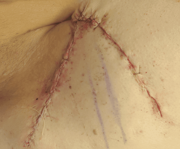
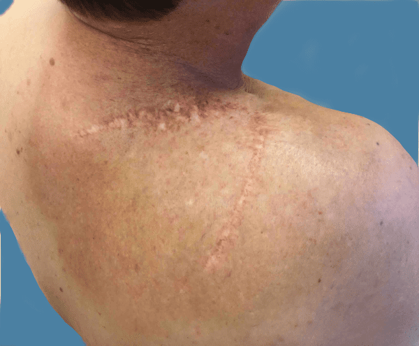
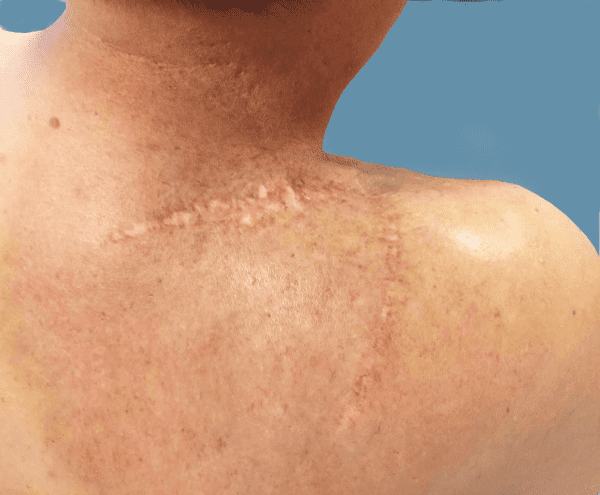
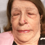
Excision Patient 01
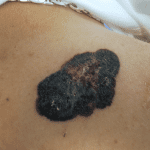
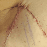
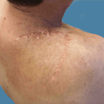
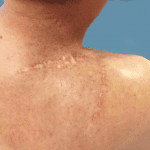
Patient with a melanoma developing from a congenital nevus on the right upper back. Patient underwent wide excision and flap closure.
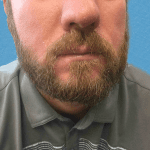
Before and After Photos - individual results may vary
Some images may be models.
Site Designed & Hosted by Plastic Surgery Studios.
Sitemap | Privacy Policy | Notice To Patients Open Payments Database
Vincent C. Hung, MD, FACS, INC. Copyright © 2025